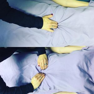
Diastasis recti in a postpartum patient, shown in the top photo below, is 3 finger width. The test being performed is a standard manual measurement whereby the patient curls their head up off the surface (as in a classic mini crunch) and the therapist measures the distance between the rectus abdominus muscles. The botttom image is showing the recti edges delineated via manual palpation, so one can confirm the findings. We also measure the inter-recti distance via rehabilitative ultrasound. The linea alba, the thick connective tissue structure that connects the left and right abdominal recti muscles, is affected by the stretching of the abdominal wall during pregnancy.

This patient had a 9 finger width diastasis recti separation originally, 3 years ago after delivery twins (she also had a singleton 2 years prior to that). We rehabilitated it successfully using an abdominal binder, prescriptive corrective exercises, neuromuscular re-education of the core, breathing patterns, and using extensive manual therapies – including visceral manipulation. She then went on to have her fourth baby. She came to see us to keep the diastasis recti during this new pregnancy at a minimum. Postpartum here she only has a 3 finger inter-recti distance. Here we prove that we can help minimize recurrence of diastasis recti if physical therapy is provided during pregnancy, especially with a patient who has a predisposition. Of note is that this patient is a die-hard runner. She jogged 4 miles several times per week through her pregnancies, despite being advised to discontinue. She also complained about terrible varicose veins of the legs which worsened with each subsequent pregnancy. Retrograde massage and lymph drainage techniques helped. She did wear compression stockings when not jogging. She has been seeing us now for 2 months and her diastasis recti has decreased to 1.5 fingers width. We are predicting a full recovery.
To read more on the topic of Diastasis Recti, click here.


